
Article: Facial Danger Zones for Botox and Fillers: A Visual Guide
Facial Danger Zones for Botox and Fillers: A Visual Guide
By Platinum Anatomy Education Team · Facial anatomy · Injection safety · Vascular anatomy
Every aesthetic injector knows the phrase danger zone. Fewer can immediately name the vessel, the depth, and the consequence — for every zone across the face.
This guide maps the primary facial injection danger zones relevant to botulinum toxin and dermal filler procedures: which vessels are at risk, at what anatomical depth, and what happens when they are compromised. It is a practical anatomical reference — a visual anchor for what you should be able to recall precisely before every treatment session.
Vascular occlusion following aesthetic injection is rare but serious. Understanding the spatial relationships between injection sites and major vessels — in three dimensions — is what separates confident, safe treatment from avoidable complications.
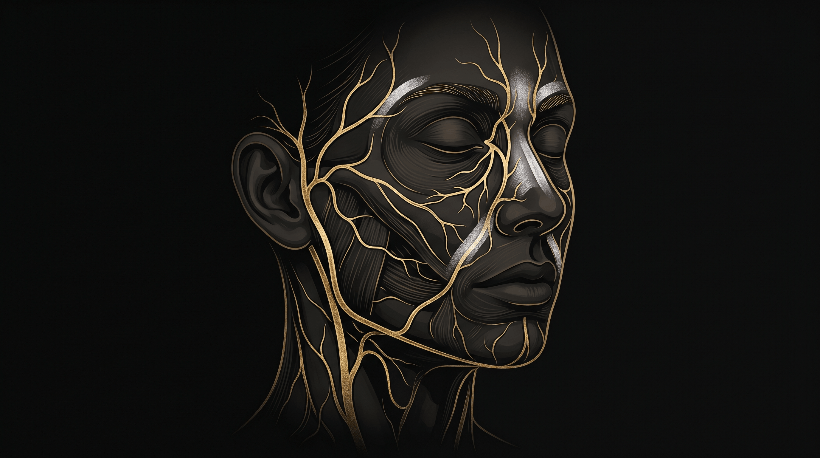
The facial vascular system — brief overview
The face is supplied primarily by the facial artery — a branch of the external carotid — and its terminal branch, the angular artery, which anastomoses with the ophthalmic artery. This anastomosis is why injections near the nose and glabella carry a risk of retrograde embolisation reaching the ophthalmic circulation.
The key arterial structures every injector must have mapped spatially before treating:
- Facial artery and angular artery — medial face, variable depth, runs beneath SMAS in lower face, becomes progressively more superficial ascending toward the nose
- Supratrochlear and supraorbital arteries — glabellar and forehead zone, exit via bony foramina, run in close proximity to standard neuromodulator injection points
- Dorsal nasal artery — nasal bridge and tip, terminal branch of the ophthalmic artery, direct retrograde embolisation corridor
- Superior and inferior labial arteries — perioral zone, run within the wet-dry vermilion border at approximately 2–3 mm depth
- Superficial temporal artery — temporal hollow, runs subcutaneously, visible and palpable in most patients
- Infraorbital artery — exits infraorbital foramen, directly relevant to tear trough and periorbital filler
- Zygomaticofacial artery — lateral orbital and cheek zone, highly variable in course and depth
The venous system largely accompanies the arterial system but is more anatomically variable. The facial vein has direct communication with the cavernous sinus via the ophthalmic veins — making the central face an area of concern for infection spread as well as vascular occlusion.
Facial injection danger zones — reference table
| Zone | Primary vessel at risk | Key risk / consequence |
|---|---|---|
| Glabellar zone | Supratrochlear + supraorbital arteries | Retrograde embolisation to ophthalmic artery — documented vision loss |
| Nasal tip & bridge | Dorsal nasal artery · Angular artery | Direct ophthalmic anastomosis — highest retrograde risk. Blindness, skin necrosis |
| Nasolabial fold | Facial artery (ascending) | Variable depth 3–12 mm — difficult to predict. Occlusion risk |
| Perioral / lip | Superior + inferior labial arteries | Intravascular injection → lip necrosis. Always aspirate. |
| Temporal hollow | Superficial temporal artery | Subcutaneous — easily hit with deep-plane filler. Blanching |
| Medial brow / glabella | Supratrochlear artery (medial) | Filler embolisation risk. Supraperiosteal approach only |
| Infraorbital / tear trough | Infraorbital artery + angular vein | Thin tissue, superficial vessels. High bruising + occlusion risk |
| Lateral orbital / temple | Zygomaticofacial artery (variable) | Variable anatomy — palpate first. Orbital fat migration risk |
| Chin / mental region | Mental artery (inferior alveolar branch) | Exits mental foramen — inject above periosteum midline only |
| Forehead lateral | Sentinel vein + frontal branch STA | Sentinel vein marks facial nerve temporal branch proximity |
High-risk zones by region — detailed breakdown
Glabellar zone — the highest-stakes injection area
The glabellar complex is the zone most consistently cited in published vascular complication reports. The supratrochlear and supraorbital arteries exit their foramina at the medial and mid-brow and run superiorly in a plane between the frontalis and the corrugator and procerus muscles — directly in the injection path for standard botulinum toxin treatment of glabellar lines.
Key risk: These vessels anastomose with the ophthalmic artery via the dorsal nasal and angular arteries. Retrograde filler injection can reach the central retinal artery. This is the mechanism behind the majority of filler-related blindness cases in the published literature.
Safe practice: Neuromodulators in the glabella carry lower vascular risk than fillers. Filler in this zone requires extreme caution: inject supraperiosteal, use micro-boluses (0.01–0.02 ml), aspirate, inject slowly, have hyaluronidase immediately available.
Nasal tip and bridge — the retrograde embolisation corridor
The dorsal nasal artery is a terminal branch of the ophthalmic artery. It descends the nasal dorsum and anastomoses with the angular artery at the nasal ala — creating a direct anatomical corridor between superficial nasal injections and the ophthalmic circulation.
Key risk: Non-surgical rhinoplasty filler injections carry the highest documented risk of blindness of any aesthetic procedure. The vessels are small (1–2 mm diameter), superficial, and surrounded by limited protective tissue.
Safe practice: Deep plane injection only — supraperiosteal on the nasal bones, supracartilaginous at the tip. Maximum 0.1 ml per injection point. Very slow injection speed. Experienced injectors only.
Nasolabial fold — the variable facial artery
The facial artery runs from the mandibular margin superiorly, crossing the nasolabial fold in a plane typically deep to the levator labii superioris. Its course is among the most anatomically variable structures in the face — position ranges from medial to lateral relative to the fold, and depth from 3 mm to 12 mm depending on the individual.
Key risk: NLF filler injections — one of the highest-volume procedures globally — carry moderate vascular risk specifically because of this variability. No surface landmark reliably predicts the artery’s position.
Safe practice: Sub-SMAS or supraperiosteal injection plane. Cannula preferred over needle (lower intravascular penetration risk). Small aliquots distributed linearly rather than single bolus. Aspirate before each bolus injection point.
Temporal hollow — the subcutaneous artery
The superficial temporal artery runs subcutaneously in the temporal region — it is among the most superficial major arteries in the face and is often visible and palpable in lean patients. Volume replacement in the temporal hollow is increasingly common in aesthetic practice, but the anatomy here is frequently undertrained.
Key risk: The STA sits in the direct path of shallow temporal filler. Intravascular injection causes blanching and occlusion. The temporal branch of the facial nerve (motor supply to frontalis) runs closely related to the sentinel vein in this zone.
Safe practice: Deep plane injection — supraperiosteal or sub-temporalis fascia — is significantly safer than subcutaneous in the temporal hollow. Always palpate for the STA before injecting. Cannula strongly preferred.
Perioral and lip — the labial arteries
The superior and inferior labial arteries run within the orbicularis oris muscle at approximately 2–3 mm depth from the mucosal surface, at or just beneath the wet-dry vermilion border. They are not reliably identifiable from the surface and anastomose freely across the midline.
Key risk: Intravascular filler injection into the labial arteries causes rapid blanching followed by necrosis of the lip mucosa or skin. Because the vessels communicate across the midline, a unilateral injection event can affect both sides of the lip.
Safe practice: Aspirate before each bolus. Use micro-aliquots of 0.05–0.1 ml maximum per injection point. Linear threading technique along the vermilion border carries lower intravascular risk than deep bolus injection.
How 3D anatomy training reduces vascular complication risk
Understanding danger zones from a diagram is a starting point. The clinical value comes from having a precise three-dimensional spatial map — knowing not just that a vessel exists, but where it runs relative to the overlying skin, the muscle beneath it, and the bony landmarks you can palpate on the patient in front of you.
A flat anatomical illustration shows you a vessel’s general course. A three-dimensional model — with colour-coded muscles and vessels at real anatomical scale — gives you the spatial depth that transfers directly to clinical practice.
Know the anatomy before you touch the patient — layer by layer.
Platinum Face II was built specifically for this type of study. It is the only 3D-printed facial anatomy training model in Europe with a complete colour-coded vascular layer — arteries in red, veins in blue — positioned in anatomically correct depth relative to the underlying muscle layer and skull base.
Rotate it. Remove the jaw. Trace the angular artery corridor from the nasal ala upward. See the labial vessels’ position within the orbicularis. Understand the superficial temporal artery’s subcutaneous path before you treat the temporal hollow on your next patient.
Pre-injection anatomy checklist
Before treating any zone with higher vascular complexity, review the following before each session:
- Can I name the primary vessel at risk in this zone — its course and approximate depth in this patient?
- Are there individual anatomical variations I need to account for — visible STA, unusually medial facial artery, prior filler in the zone?
- What injection plane am I using — and is it the safest available plane for this specific zone?
- What is my bolus volume — and is it the minimum clinically necessary?
- Am I using the safest delivery method — cannula vs needle — for this zone and this patient?
- Is hyaluronidase immediately accessible in the room?
- Do I know the early signs of vascular occlusion and my management protocol for the first 60 seconds?
Summary
Facial injection danger zone knowledge is not advanced anatomy — it is the baseline for every aesthetic injector regardless of experience level. The vascular anatomy of the face is complex, individually variable, and forgiving of very little.
The zones most consistently associated with serious complications are the glabellar complex, the nasal dorsum, the nasolabial fold, the temporal hollow, and the perioral region. Every one of these should be studied in three dimensions before treating.
Study facial vascular anatomy in 3D — Platinum Face II
Platinum Face II is the only 3D-printed facial anatomy training model in Europe with a complete colour-coded vascular system — arteries, veins and danger zones mapped at real anatomical scale.
→ View Platinum Face II — from €725
→ Compare all Platinum Anatomy models
→ For Academies & Distributors — volume pricing from 3 units
Related articles — coming soon
- How to Choose a Facial Anatomy Model for Injection Training
- Facial Muscles of Expression: Anatomy Every Injector Must Know
- Why Every Aesthetic Injector Needs a 3D-Printed Anatomy Model
